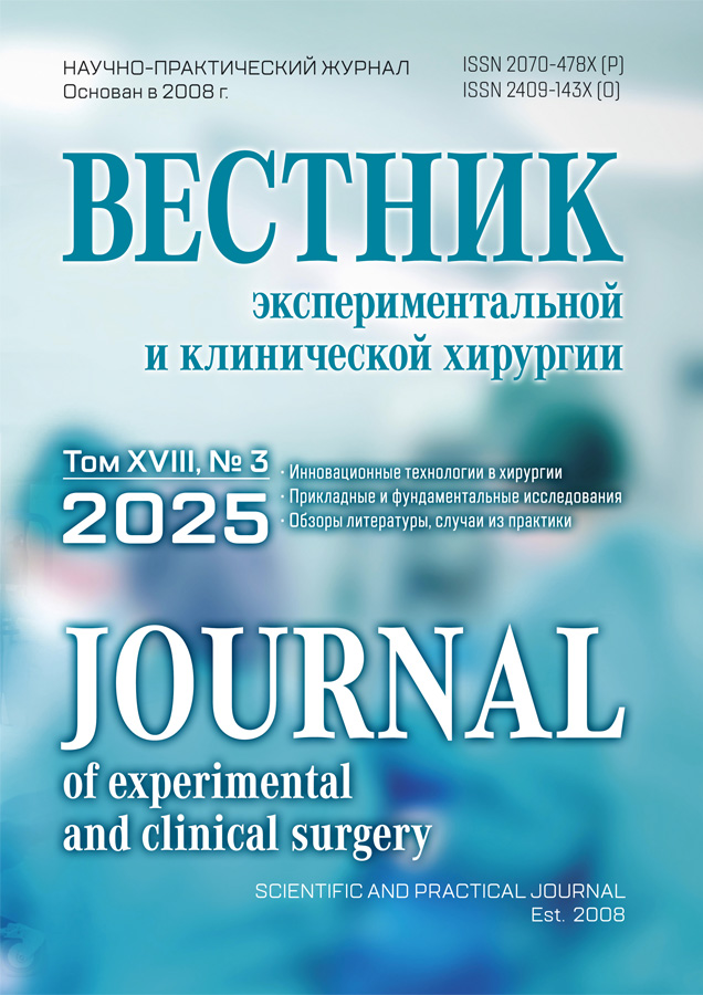
Journal of Experimental and Clinical Surgery
Scientific and practical journal "Journal of Experimental and Clinical Surgery" is established by the Institute of Surgical Infection of N.N. Burdenko Voronezh State Medical Academy in 2008.
The journal publishes original articles on clinical and experimental studies, literature reviews, brief reports on clinical observations, information on innovations, inventions and innovative projects, new methods of diagnosis and treatment, materials on the history of departments, clinics and surgical hospitals.
Current Issue
Vol 18, No 3 (2025)
Original articles
Pancreaticoduodenectomy in a soft pancreas
Abstract
Aim. To evaluate the effects of different techniques of the pancreaticoduodenectomy (PD) on the frequency and severity of postoperative complications and mortality in patients with a "soft" pancreas.
Material and methods. The results of 141 PDs were evaluated (76 were exposed the conventional surgery and 65 were exposed to the expanded volume of pancreatic resection, considering angioarchitectonics). In all cases, the pancreas was "soft", which was confirmed by morphological examination. The arterial architectonics of the cephalocervical segment of the pancreas was studied in the anatomical part of the study on 94 preparations, as well as clinical and radiological examination in 62 people.
Results. In patients with "soft" pancreas, an increased volume of resection during PD, considering arterial architectonics in the cervical region, allowed for a slight improvement in the immediate results of the operation: the number of specific postoperative complications decreased from 51.3% to 43.1%, mortality decreased from 7.9% to 4.6%. The probability of a postoperative pancreatic fistula (POPF) increased in cases where the dorsal pancreatic artery was not a branch of the splenic artery. The formation of an anastomosis with a pancreatic stump on an isolated loop and the preservation of the gastrointestinal ligament at the stage of mobilization reduced the aggressiveness of postoperative complications.
Conclusion. The expanded volume of pancreatic resection to ensure adequate blood supply of the stump of the organ appears to be an effective option only in cases of dorsal pancreatic artery withdrawal from the splenic artery. It is not possible to avoid ischemia of the pancreatic stump in other variants of the arterial anatomy of this vessel. In case of POPF, reconstruction involving two loops of the jejunum can reduce its aggressiveness. Mobilization of the complex with preservation of the gastrointestinal ligament facilitates the identification of the superior mesenteric vein and reduces the frequency of gastrostasis in the postoperative period.
 177-187


Undifferentiated connective tissue dysplasia as a risk factor for recurrence of postoperative ventral hernia
Abstract
Backgraund. Despite the development of surgical techniques, the recurrence rate of postoperative hernias (PH) remains high. There is increasing evidence in the literature on the role of connective tissue pathology in the development and recurrence of postoperative ventral hernias
Aim. Establish the relationship between the severity of connective tissue dysplasia (CTD) and the results of plastic PH also identify the correspondence between the phenotypic and morphological signs of CTD in patients with PH.
Materials and methods. The study was conducted on the basis of the "Petrovsky National Research Centre of Surgery" and GBUZ "GCB No. 52 DZM" from 2018 to 2024. The main group consisted of 91 patients with postoperative hernia, the control group consisted of 10 patients who underwent surgery with a history of laparotomy, but without the formation of PH. The groups are identical in age, gender ratio, and comorbidity. 25 plastic surgeries were performed with local tissues and 66 with the use of a mesh prosthesis, morphometric studies of aponeurosis for the ratio of type 1 and type 3 collagen were performed in 50 patients. All patients were assessed for the severity of external signs of connective tissue dysplasia using the Luzgina-Shkurupiya method. The reliability of differences between the groups in the number of hernia recurrences was assessed for each group by the coefficient of statistically significant differences calculated using the Pearson's χ2 criterion. The ratio of collagen types was calculated by analyzing micrographs based on a color histogram in Adobe Photoshop with the calculation of the average number of pixels of different colors for the entire preparation: type 1 collagen - red spectrum, type 3 – green. Quantitative indicators were evaluated for compliance with the normal distribution using the Shapiro-Wilk criterion. Quantitative indicators, the sample distribution of which corresponded to the normal, were described using arithmetic averages (M) and standard deviations (SD). The boundaries of the 95% confidence interval (95% CI) were indicated as a measure of representativeness for the mean values. A comparison of three or more groups by a quantitative indicator, the distribution of which in each group corresponded to the normal one, was performed using a one-factor analysis of variance, aposteriori comparisons were carried out using the Tukey criterion (assuming equality of variances). The differences were considered statistically significant at p < 0.05.
Results. When comparing patients with PH and dysplasia of mild and severe severity, statistically significant differences in the number of relapses were revealed, including with mesh plastics (P<0.05). Statistically significant (P<0.05) differences in the number of pixels were found, reflecting the ratio of type 1 and type 3 collagen, in the direction of an increase in type 3 collagen with an increase in the severity of connective tissue dysplasia
Conclusions. It was found that the probability of PH recurrence significantly increases with increasing severity of CTD, therefore, assessment of the severity of CTD is an important element in the treatment of patients with PH. This makes it possible to prevent the recurrence of PH by choosing the most appropriate surgical technique. Diagnosis of CTD is carried out by phenotypic and morphological methods. The study proved that there is a direct relationship between the phenotypic manifestations of CTD and its morphological features. Therefore, both methods can be adequately applied to assess the severity of CTD.
 188-194


Cases from practice
Combined pancreatic solid-pseudo-papillary tumor and spleen lesions: diagnosis complexities in determining treatment tactics
Abstract
Solid pseudopapillary tumors (SPTs) are rare neoplasms, which account for less than 2% of exocrine pancreatic tumors. These are low-grade epithelial malignancies. Currently, there are no data on SPT metastasis to the spleen in the literature. However, due to the rare SPT incidence and the ongoing accumulation and data analysis on these type of tumors globally, the presence of a combined cystic-solid structure lesion in the spleen does not allow us to exclude such a situation.
Two clinical cases of SPT combined with focal spleen lesions are presented. The first one is with a cystic lesion verified as cavernous spleen lymphangioma according to the morphology of the removed lesion. Despite the fact that the splenic lesions included in the differential series during examination were benign, the size of the lesion and the risk of its rupture demonstrated the advisability of spleen resection performing.
The second one is combined with multiple spleen hemangiomas verified by preoperative MSCT, which allowed performing a sparing operation - median resection (tumor enucleation) of the pancreatic head.
Thus, а thorough examination of patients with combined focal lesions, one of which being SPT, a tumor with malignant potential, allows us to clearly determine the entire volume of the lesion and determine the correct surgical tactics.
 195-207


A giant thymolipoma of the anterior mediastinum extending into the right hemithorax: a rare clinical case
Abstract
Thymolipoma is a rare benign neoplasm of the chest cavity. Due to the long asymptomatic course, this pathology is detected in patients accidentally or when the tumor reaches gigantic sizes, when the compression syndrome of the chest organs and mediastinum manifests. Diagnosis of this tumor is difficult due to the variety of neoplasms of the anterior mediastinum. Detecting tumor tissue based to CT data is not difficult. However, verification of the diagnosis causes a number of difficulties due to the low information content of the obtained biopsy, which demonstrates only the presence of fatty tissue. When determining the exact diagnosis of "thymolipoma", the main treatment option is surgical removal of the tumor. The surgical approach should be chosen taking into account its size and topographic characteristics. This article presents a rare clinical observation of a patient with a giant thymolipoma of the anterior mediastinum with extension to the right hemithorax, who underwent radical surgical treatment through a sternotomy approach.
 208-212


Boerhaave syndrome associated with portal hypertension with varicose veins of the esophagus
Abstract
Spontaneous rupture of the esophagus is a disease that directly threatens the patient's life. One of the earliest descriptions of the beginning of the XVIII century belongs to the Dutch physician H. Burchaave. A clinical case of treatment of a female patient with spontaneous rupture of the esophagus (Burhave syndrome) against the background of cirrhosis of the liver and significant varicose veins of the esophagus is presented. When using extracavitary vacuum therapy, rapid epithelialization of the esophageal rupture site was noted, followed by regression of the cavity in the mediastinum. This could be facilitated by a specific thickening of the wall caused by varicose veins of the esophagus.
Subsequently, on the 12th day of follow-up, the patient developed massive bleeding from varicose veins of the cardiac stomach, and therefore underwent emergency surgery. The fatal outcome was due to the development of postoperative complications against the background of severe initial pathology. According to autopsy data, complete healing of the esophageal defect and resolution of mediastinitis were noted.
Thus, in such a serious life-threatening condition as spontaneous rupture of the esophagus, varicose veins can play the role of a protective factor in the form of temporary tamponade of the injury zone and prevent the lightning-fast development of mediastinitis.
 213-217


Review of literature
Bariatric surgery in patients with first-degree obesity: is the surgical approach reasonable?
Abstract
The widespread progression of obesity, the insufficient effectiveness and stability of conservative approaches in its treatment and the corresponding development of bariatric surgery drive the need to study the potential and safety of performing surgery in patients with first-degree obesity. The article analyzes the literature data of research results presenting the experience of bariatric surgery application in patients with a body mass index (BMI) equal to 30-34 kg/m2, comparing the outcomes of surgery and the use of conservative treatment options for obesity and evaluating the effectiveness and safety of various types of bariatric surgery in this group of patients.
Due to controversy of the issue currently being discussed, the aim of the study was to assess the safety, effectiveness, and reasonability of bariatric interventions in patients with a relatively small excess weight based on available literature data.
 217-224


